×

We have detected your country as:
Please click here to go to the USA website or select another country from the dropdown list.
We have detected your country as:
Please click here to go to the USA website or select another country from the dropdown list.


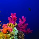
Photo: sergemi/shutterstock.com
We know that human-induced environmental changes are responsible for coral bleaching, disease, and infertility. Loss of the world’s stony coral reefs—up to 30% in the next 30 years, according to some estimates—will mean loss of their services, including sequestering some 70-90 million tons of carbon each year and supporting enormous marine biodiversity. Yet despite many advances, we are still far from understanding the causes and processes contributing to the corals’ demise.
Weizmann Institute researchers have developed a new experimental platform for studying coral biology at microscale resolutions, which is already providing new insights into this complex problem.
The tiny—often less than a millimeter in diameter—animals that build coral reefs create a thin layer of living tissue surrounding the calcium-based skeleton. These animals live in symbiosis with single-celled, photosynthetic algae that provide nutrients and oxygen in return for carbon dioxide and shelter. “In order to understand what happens during bleaching, when this symbiosis is broken,” says Dr. Assaf Vardi, “we need to understand what happens to these organisms at the cellular and molecular levels under various conditions.”
Vardi and his team created a system they call “coral on a chip.” For the first time, the scientists were able to examine living coral polyps in the lab, under highly controlled conditions based on microfluidics technology.
Taking a small piece of coral, Vardi and his team induced stressful conditions—in this case by increasing salt content—which caused the corals to release polyps, a process sometimes referred to as “polyp bail-out.” Settling the bailed-out polyps into prefabricated microfluidic wells, the scientists were able to observe how miniature coral colonies called “micropropagates” grow and behave in different conditions.
Using their system, the team recorded, for the first time, the growth of individual aragonite crystals—the basic building blocks of the coral skeleton. The group was also able to directly visualize the initiation of coral disease, pointing to a little-known path of infection. Subjecting coral micropropagates to high light intensities enabled the team to follow the elimination of the symbiotic algae, one cell at a time. Vardi said, “Many corals are running out of time; it is crucial to know how our actions are affecting their survival, and how they affect ours.”
Source: Excerpt of an article, Israel Ministry of Foreign Affairs


All logos and trademarks in this site are property of their respective owner. All other materials are property of Bridges for Peace. Copyright © 2024.
Website Site Design by J-Town Internet Services Ltd. - Based in Jerusalem and Serving the World.
